
Understanding Arthritis: How Thermography Offers Insight and Relief

Arthritis is a common condition that affects millions of people worldwide, causing pain, and stiffness in the joints. While X-rays and MRIs are valuable, they may not always provide a comprehensive understanding of arthritis-related inflammation. Thermography, a non-invasive imaging technique, offers a unique perspective on arthritis by revealing areas of inflammation. In this blog, we’ll explore arthritis, how thermography works, and its potential benefits in managing this chronic condition.

Understanding Arthritis:

Arthritis is a term used to describe inflammation, which can result in pain, swelling, stiffness, and reduced range of motion. There are many types of arthritis, including osteoarthritis (the most common form), rheumatoid arthritis, psoriatic arthritis, and gout, each with its unique causes and symptoms. While arthritis is typically associated with aging, it can affect people of all ages and lifestyles.

The specific characteristics and underlying mechanisms of inflammation can vary depending on the type of arthritis and individual factors.

Traditional Diagnostic Methods:

Traditional diagnostic methods for arthritis, such as X-rays and MRIs often involve radiation exposure which may not be suitable for all individuals.

How Thermography Works:

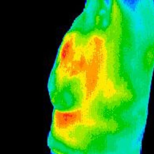
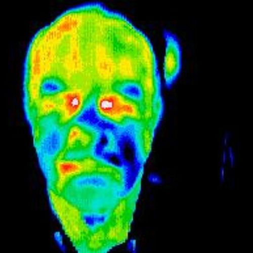
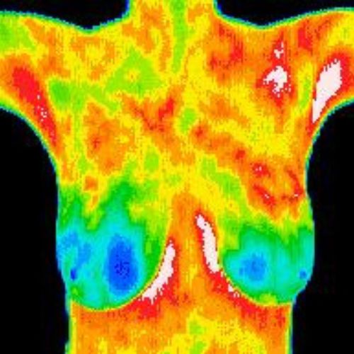
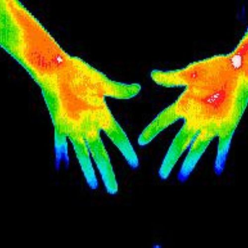
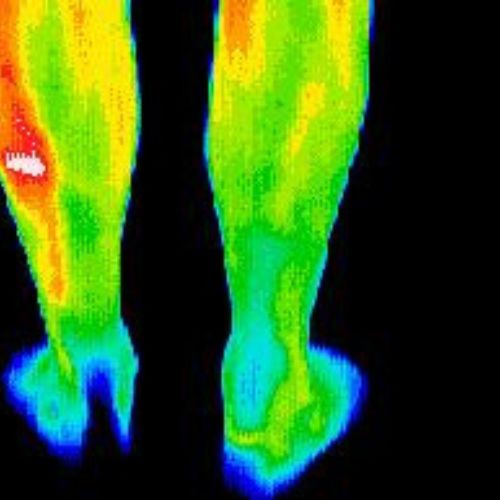
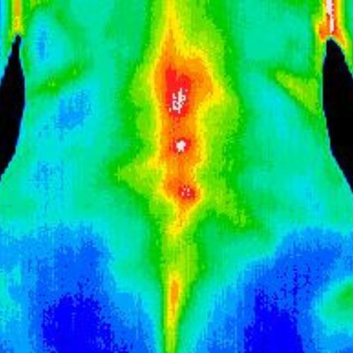
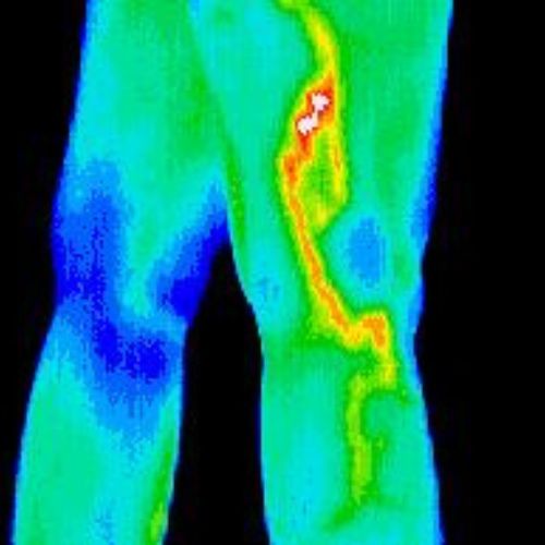
Thermography, also known as infrared imaging, is a non-invasive imaging technique that captures heat patterns. By detecting variations in temperature, thermography can identify areas of inflammation and increased blood flow, which are common indicators of arthritis. During a scan, a thermal camera records infrared radiation emitted by the body and generates a visual map of temperature distribution.

The Potential Benefits of Thermography in Arthritis Management:

  1. Early Detection: Thermography may detect signs of arthritis-related inflammation in its early stages, allowing for prompt intervention and treatment.
  2. Non-Invasive: Unlike traditional imaging methods, thermography is non-invasive and does not involve radiation or contrast agents, making it safe for repeated use and suitable for individuals with contraindications to other imaging modalities.
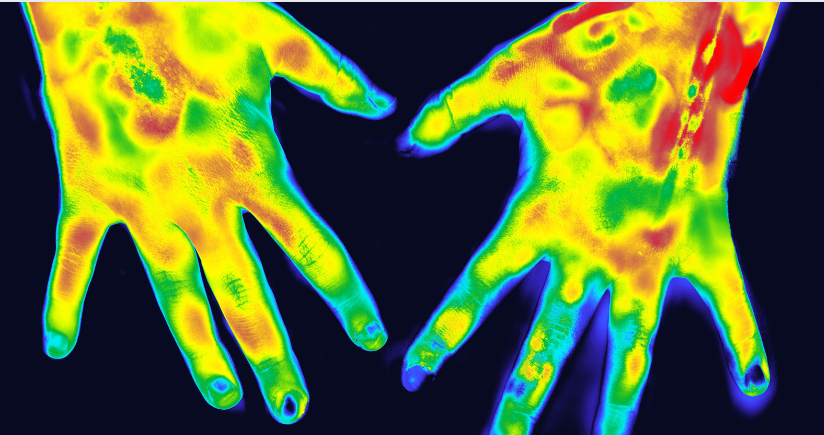
  3. Comprehensive Assessment: Thermography provides a holistic view of arthritis-related inflammation by capturing heat patterns across multiple joints simultaneously.
  4. Monitoring Disease Progression: Thermography can be used to monitor changes in inflammation over time, providing valuable information for assessing treatment and disease progression.
  5. Personalized Treatment Planning: By identifying areas of inflammation, thermography may inform personalized treatment plans tailored to address individual needs and target specific areas.

I.V. therapy for Inflammation:

Intravenous (IV) therapy, often referred to as IV infusion therapy, has gained popularity as a potential treatment for inflammation. IV therapy involves administering fluids, vitamins, minerals, and other nutrients directly into the bloodstream.

Anti-inflammatory Ingredients:

Some IV therapy formulations may include ingredients known for their anti-inflammatory properties, such as vitamin C, vitamin B complex, glutathione, magnesium, and zinc. These nutrients are believed to help reduce inflammation and support the body’s natural healing processes.

  • Safety and Sterility: IV therapy should be performed in a sterile clinical setting by trained professionals to minimize complications.

Arthritis can significantly impact quality of life, but early detection and comprehensive management are key to minimizing its effects. Thermography offers a non-invasive and holistic approach to assessing arthritis-related inflammation. Thermography holds promise as a valuable tool in the multidisciplinary approach to arthritis care.

If you are living with arthritis, consider exploring thermography as part of your comprehensive treatment plan. Consult with a qualified healthcare practitioner to learn more about how thermography may benefit your overall health.

Breast Health Screening: Thermography vs Mammogram

Breast health is a paramount concern for women worldwide, with early detection being key to successful outcomes in breast cancer management. While mammography remains the gold standard for breast cancer screening, emerging technologies offer complementary approaches that enhance detection and monitoring. One such innovation is breast thermography, a non-invasive imaging technique that holds promise for early detection and risk assessment. In this blog post, we explore the principles of breast thermography, its benefits, and its role in modern breast health management. So, let’s help you decide Thermography vs Mammogram.

Breast Thermography:

An Alternative Approach Breast thermography is a non-invasive imaging technique that detects temperature variations in breast tissue using infrared technology. Areas of increased metabolic activity, such as cancerous or precancerous lesions, exhibit higher temperatures, which are visualized as color-coded images.

Benefits of Breast Thermography:

  1. Non-Invasive: Unlike mammography, which involves breast compression and exposure to ionizing radiation, thermography is non-invasive and painless. It poses no risk of radiation exposure or tissue damage, making it suitable for repeated screenings and for women with breast implants or dense breast tissue.
  2. Early Detection Potential: Thermography can detect physiological changes in breast tissue years before they become apparent on mammograms or clinical exams, offering potential for early intervention and improved outcomes.
  3. Dynamic Monitoring: Breast thermography allows for dynamic monitoring of breast health over time, facilitating longitudinal assessments of changes in temperature patterns and enabling personalized risk management.
  4. Dynamic Monitoring: Breast thermography allows for dynamic monitoring of breast health over time, facilitating longitudinal assessments of changes in temperature patterns. Regular thermographic screenings can help detect subtle alterations in breast physiology, guiding early intervention and personalized risk management plans.

Understanding Breast Thermography:

Breast thermography, also known as infrared thermography or thermal imaging of the breast, utilizes infrared technology to detect temperature variations in breast tissue. These temperature differences reflect metabolic activity and blood flow patterns, which can be indicative of underlying breast pathology, including cancerous and precancerous lesions.

Unlike mammography, which relies on X-rays to visualize breast tissue density, thermography captures heat emissions from the body’s surface. Areas of increased metabolic activity, such as tumors or inflammation, exhibit higher temperatures, which are detected by thermographic cameras and translated into color-coded images for analysis.

Integrative Approach to Breast Health:

While breast thermography offers promise as a complementary screening tool, it is not intended to replace mammography or clinical breast exams. Rather, it serves as a valuable adjunct to existing breast health protocols, providing additional information for comprehensive risk assessment and early detection.

A holistic approach to breast health incorporates multiple modalities, including mammography, ultrasound, magnetic resonance imaging (MRI), and clinical breast exams, tailored to individual risk factors and preferences. Breast thermography complements these modalities by offering unique insights into breast physiology and enhancing detection sensitivity, particularly in cases of dense breast tissue or in women at higher risk for breast cancer.

Breast thermography represents a groundbreaking advancement in breast health monitoring, offering early detection, risk assessment, and dynamic monitoring capabilities. While it is not a standalone diagnostic tool, its non-invasive nature, early detection potential, and ability to assess physiological changes make it a valuable addition to the armamentarium of breast cancer screening modalities.

As research and technology continue to evolve, the integration of breast thermography into standard breast health protocols holds promise for improving detection rates, reducing unnecessary interventions, and ultimately saving lives. By embracing a comprehensive and integrative approach to breast health management, we can empower women to take proactive steps towards early detection, prevention, and optimal breast health outcomes.

Discover if Breast Thermography is Right for You: Schedule Your Appointment with One of Our Health Practitioners Today!

Women’s Thermography Breast Screenings

Breast cancer is most curable when it’s detected in the early stages, so women are eager to find tools to help them in their battle against cancer. 

Women often schedule Thermography, in addition to annual mammograms, to improve their chances of detecting changes in breast tissue.

Serenity is dedicated to breast cancer awareness

We hold breast cancer awareness events regularly. The foundation of our breast cancer awareness protocol is Thermography.

We advocate this harmless technology not only for breast cancer but for the detection of numerous other diseases including heart disease, digestive disorders, arthritis, and diabetes complications.

Breast thermography, when used as part of a woman’s breast cancer awareness program, has many benefits

The benefits are:

  • It helps identify patients who may be at high risk for developing breast cancer – male or female.
  • Breast thermography can increase the effective use of mammography by providing a clinical marker for physicians and radiologists. Thermography identifies a particular breast area that needs closer examination.
  • It benefits younger women who may have dense breast tissue or who may not be receiving mammograms due to current guidelines.
  • No radiation is involved.
  • No contact with your breasts.
  • No risk of torque compression.
  • It’s a low-cost screening procedure.

How accurate is breast screening thermography?

Serenity recommends Thermography or DITI (Digital Infrared Thermal Imaging) in conjunction with mammography. Thermal imaging is non-invasive and is 90% accurate for cancer detection (vs. 75% for traditional mammography). Thermography detects cancer up to 10 years sooner than conventional mammography.

Thermography gives the practitioner a color map of the body’s temperature. Abnormal physiology presents in different colors within the scan. 

Is Thermography as good as mammography?

Breast thermography is an excellent screening tool that women can add to their annual mammograms. Thermography digital imaging technology, has two distinct advantages over other breast cancer screening tools:

1. It is non-invasive

2. It does not emit any harmful radiation

The procedure lasts about fifteen minutes, and no equipment touches the breast. 

Digital infrared thermal imaging effectively detects potential problem areas in breast tissue that may indicate malignancy in the early stages because inflammation is visible prior to disease as the body begins gearing up to fight unwanted cells.

Breast thermography is an FDA-approved procedure that can help visualize and quantify changes in skin temperature. 

If breast thermography detects any suspicious findings, women can alert their physician so that they can pursue further testing and treatment. 

Breast thermography does not take the place of mammography when that test is deemed medically appropriate.

Don’t delay. Book your breast screening now here

How does breast thermography work?

Breast thermography employs digital infrared thermal imaging technology. 

An infrared scanning device, which does not touch the body, measures the skin surface temperature of the breasts. 

The device converts infrared heat that the skin naturally emits into electrical impulses projected on a monitor. As a result, breast thermography maps the body temperature. 

The spectrum of colors on the monitor indicates the amount of infrared heat emitted from the breasts’ surface.

Chest and breast screening of 4 different women and men showing colorful diseases in a thermogram.

Are you under 50? Younger women rely on Breast Thermography screening

Younger women, ages 30 to 50, often have dense breast tissue. Dense breast tissue can make it difficult for a screening mammogram to detect breast cancer. 

In addition, many physicians do not recommend mammograms for women under 50. So, breast thermography can serve as an early detection and monitoring tool for young women with a genetic predisposition to the disease.

What is the cost of thermography for breasts?

Most of our patients receive a full body Thermography scan because it helps to uncover hidden potential health issues so we can prescribe preventative health measures. 

The technology is ideal for preventative medicine and to identify the underlying cause of your regionalized inflammation, pain or swelling.Breast scans are often coupled with Lymph Enhancement Therapy as an integrative protocol. Depending on the requirements, scans range from around $350 and up, but prices depend on the body surface areas needed. The best way to get an estimate is to call our front desk or drop us an email.

Serenity’s Thermography expertise is available to you in Waukesha, WI

Serenity Health Care Center has the equipment and knowledge available to give our patients cutting-edge Thermography scans. 

Although patients can expect results from their test in 2-3 days, we can sometimes expedite within 24 hours if we see something that needs an urgent diagnosis.

Book a Thermography Scan here

Did you know that it takes 8-10 years of training to read results accurately?

from top left a thermogram of normal breasts, followed by an inflammatory breast cancer, followed by estrogen dominance thermogram, followed by an Inmune dysfunction thermogram, followed by a photo of male breast cancer and n the bottom left a thermogram of shoulder pain.

Serenity Health Care Center works with certified experts to perform this excellent diagnostic, and prognostic testing with trustworthy result interpretations.

At our functional medicine clinic near Milwaukee, breast thermography allows us to:

  • Detect and monitor abnormal physiology
  • Establish risk factors for developing breast disorders
  • Detect early indications that breast cancer may be present

If you want to know more about Thermography and its benefits visit our Medicine Thermography page

Call 262.522.8640 today to schedule your Thermogram scan with the professionals at Serenity Health Care Center in Waukesha

We can understand and see your pain with Inflammation Thermography

woman's knee photo showing a red halo as if it was painful due to inflammation

Inflammation is part of your natural healing process in response to infections and injuries. Your inflammatory response is a healthy way for your body to dispose of pathogens. Inflammation also isolates the injury by preventing adjacent tissues from damage. Inflammation flushes and cleans because the process helps your cells get rid of debris. 

In other words, your body repairs itself with inflammation – which is a good thing. 

Nevertheless, chronic low-grade inflammation is scientifically proven to cause cancer, heart disease, type 2 diabetes, and other deadly diseases. Inflammation involves chemical reactions and cellular function, and prolonged artery, capillary, and vein dilatation can tax your system and cause long-term health concerns.

By allowing inflammation to go unchecked, your entire body kicks into high gear to help combat illness. Unfortunately, sometimes your body goes overboard in the process and weakens over time – which is when more complex health problems arise. 

So, inflammation is not only an indicator of a health abnormality but also the cause of disease. Serenity wants to help you identify and control inflammation.

What is Thermography for Inflammation?

Using our Thermography camera detector, this technique precisely records the energy emitted in the infrared spectrum. Thermography uses infrared radiation (photons) emitted by your body into electrical signals that form a digital image on a monitor.

Even at a distance and without any direct contact, Serenity can measure the temperature of your inflamed area using the infrared detector because areas with increased blood flow indicate inflammation. Inflammation appears in red, while areas with little blood flow activity show in cooler colors. 

So, if we can see your health problem, we can help get it under control – and sometimes even reverse illness!

What can Thermography detect based on your inflammation levels?

Thermography not only detects illness based on inflammation but also measures the severity of the disease process. 

Inflammation is visible on Thermography scans with a variety of health concerns, including:

If you are a woman, Serenity is particularly interested in helping you fight against the risk of Breast Cancer. 

Thermography is helpful in the detection of Breast Cancer. It offers many benefits, including detecting Breast Cancer 7-10 years earlier than a mammogram. This is because mammograms are not prescribed to younger women and also because inflammation begins in the early stages of Breast Cancer.

Continue reading our blog to learn more about Breast Thermography Screening

Can Thermography detect infection?

Thermography can detect viral and bacterial infections because these invaders activate your immune system. Your immune system uses inflammatory cells and cytokines (substances that stimulate more inflammatory cells).  The job of these cells is to trap bacterial or viral intruders and activate your natural healing powers. These cells can cause pain, swelling, bruising, or redness – all of which are visible on a Thermography scan.

Why is Thermography used in medical diagnosis?

The temperature of a body or object correlates to the amount of infrared radiation emitted. Infrared radiation is invisible to the human eye. 

Thermography is expanding in diverse sectors such as engineering, rescue, meteorology, astronomy, public safety, electronics, meteorology, industry, and ecological analysis. 

Medical applications for this technology are making great strides; nevertheless, Serenity has been using Thermography for over a decade for the diagnosis and prognosis of our patients.

In case of infection and inflammation, the increase in temperature is clearly visible. However, depending on the severity of the lesion and the length of time of the problem, there may be a decrease in local temperature. This is because the body has given up its attempt to heal, or the lesion is so ingrained that no blood reaches the area anymore.

Continue reading our blog to learn more about: What is Medicine Thermography?

How can Serenity tell what your Inflammation Thermography results mean?

Body heat constantly dissipates from the skin by radiation, convection, conduction, or evaporation. Pathologies produce variations in blood circulation, which affect skin temperature. Due to these heat losses, the skin temperature is 5 °C lower than the internal body temperature. Thermographic imaging quantifies the skin’s surface temperature but also senses that of the deep tissues.

One crucial factor Serenity detects is symmetry. Both sides of a healthy body tend to emit similar color mapping. A marked difference requires further investigation and testing. 

Our experienced staff knows what typical heat emissions for organs look like. Trained professionals easily detect abnormalities.

To read Thermography results, certified professionals need at least ten years of training!

Serenity can even detect hidden Inflammation to avoid future health problems

Thermography reveals the primary area of injury and helps recognize secondary points of pain too. 

Secondary inflammation may not always appear in a clinical examination – but it also needs to be addressed with Integrative Medicine protocols. 

A significant advantage of this technique is that it allows early diagnosis of pathologies such as tendinitis or degenerative joint disease. 

Thermographic alterations are frequently evident before physical ones. 

Thermography can detect lesions 1 or 2 weeks before limps or pain manifest, which athletes often leverage the technology.

Once treatment begins, thermography can monitor progress. Your provider’s prescribed holistic treatment should produce the desired circulatory change. 

For example, if visible red hot inflammation is present with your baseline Thermography, then in subsequent scans, a decrease in temperature to normal levels will prove your healing progress.

In Summary

Serenity patients like you benefit from our experience with Thermography. We’ve provided this state-of-the-art technology to our patients for over a decade.

Thermography imaging technology, provides our skilled practitioners with valuable information that helps them accurately diagnose your wellness concerns and symptoms.

Book your Thermography Scan today!

Thermography Center

Medical Thermography is a non-invasive diagnostic technique that allows the examiner to visualize and quantify changes in skin surface temperature.

Heat patterns indicate inflammation. Inflammation is a precursor to disease in the body. Early detection of inflammation is critical – you can prevent or intervene in the process before it becomes a disease. 

In addition, measuring inflammation consistently over time can also be very effective for detecting the status and severity of a medical condition.

What is Thermography for Inflammation?

Using our Thermography camera detector, this technique precisely records the energy emitted in the infrared spectrum. Thermography uses infrared radiation (photons) emitted by your body into electrical signals that form a digital image on a monitor.

Even at a distance and without any direct contact, Serenity can measure the temperature of your inflamed area using the infrared detector because areas with increased blood flow indicate inflammation. Inflammation appears in red, while areas with little blood flow activity show in cooler colors. 

So, if we can see your health problem, we can help get it under control – and sometimes even reverse illness!

More About Thermography

Explore our growing collection of articles and services related to Thermography.

In Summary

Serenity patients like you benefit from our experience with Thermography. We’ve provided this state-of-the-art technology to our patients for over a decade.

Thermography imaging technology, provides our skilled practitioners with valuable information that helps them accurately diagnose your wellness concerns and symptoms.

Book your Thermography Scan today!

Continue reading

Medical Thermography

Medical Thermography is a non-invasive diagnostic technique that allows the examiner to visualize and quantify changes in skin surface temperature.

Heat patterns indicate inflammation. Inflammation is a precursor to disease in the body.

Early detection of inflammation is critical – you can prevent or intervene in the process before it becomes a disease. 

In addition, measuring inflammation consistently over time can also be very effective for detecting the status and severity of a medical condition.

How is Medical Thermography testing done?

An infrared scanning device converts infrared heat emitted from the skin surface into electrical impulses visualized in color (16 different pixels) on a monitor. 

This visual image graphically maps the body temperature and is referred to as a Thermogram.

This 15 to 30-minute valuable procedure helps alert your Integrative Medicine practitioner to the possibility of underlying breast disease and other chronic illnesses, including heart disease, and digestive disorders.

What does a Thermography scan show exactly?

Pain in the body shows up as Hyperthermia (hot spots: red and white) or Hypothermia (cold spots: blue, purple, and black), indicating to the client and practitioner a vascular or nerve problem within the body.

The spectrum of colors indicates an increase or decrease in the amount of infrared heat emitted from your body’s surface.

Since the average body has a high degree of thermal symmetry, subtle abnormal temperature asymmetries can be easily identified.

What diseases can Thermography detect?


1. Breast and chest Cancer’s early signs:

The Medical Thermography test improves your chances of detecting fast-growing, active tumors in the intervals between mammographic screenings (or when mammography is not indicated by screening guidelines for women less than 50 years old).

Your breast thermograms are kept on record and form a baseline for all future routine evaluations.

Breast thermography does not take the place of mammography when a mammogram is deemed medically appropriate.

2. The mouth is the source of many worrisome ailments:

TMJ, infected root canals, and gum disease can be revealed through Thermography scans. As well as pinpointing the source of pain, Thermography can also monitor the healing process.

3. Heart disease, stroke, and high blood pressure:

Used as a preventative tool, Thermography can be an excellent resource for vascular problems and carotid arteries. 

Thermography uncovers reduced blood flow caused by hardened arteries – a symptom of atherosclerosis. 

This technology avoids radiation, risk, dye injections, and drugs so that our practitioners can prescribe reparation protocols before it is too late. If caught soon enough, harmful fibrous tissues and plaque can be restored. 

Diet, lifestyle adjustment, supplements, and chelation detoxification go a long way toward avoiding surgery or stroke.

4. Arthritis:

Duke University has determined that Thermography identifies minimal temperature changes in joints which is the first sign of the inflammation accompanying arthritis. 

The earliest inflammatory phase of arthritis shows clearly as heat; however, as the disease worsens, the joint temperature cools significantly because of joint disuse and lack of blood supply. 

Therefore, monitoring arthritis with Thermography can help determine if your treatment is working to halt further deterioration of joint movement.

5. Diabetes:

If you have diabetes, our goal is to avoid dangerous complications. 

Thermography is a  prognostic and diagnostic tool for type 2 diabetes. It helps in the early detection of neuropathy and vascular disorder, and diabetic foot. 

6. Pain syndrome from injuries:

Diagnosing pain syndromes is one of the lesser-known benefits of Thermography. 

If you develop this difficult-to-diagnose syndrome, you may suffer from pain for six months or longer after an injury. Thermography helps diagnose sports injuries, sprains, inflamed tendons, and pain syndromes so that you can receive treatment.

7. Cancer:

Cancer and malignant tumors show on Medicine Thermography scans. 

We can not guarantee that all cancer can be detected during your appointment. However, if there are cancer indications, we will conduct further tests to confirm the abnormality visible on the color digital image.

Is thermography painful?

No. Thermography is painless. It is like taking a photograph.

In summary

Medical Thermography’s role is to help with the early detection and monitoring of abnormal physiology and establishing risk factors for the developing or existing disease. In addition, when used with other diagnostic procedures, it assists in providing the best possible holistic health evaluation.

Book your Medicine Thermography scan today

Benefits of Thermography

Learn the benefits of thermography

Learn the benefits of thermography

We want you to know about the benefits of thermography, a technology we offer at our functional medicine clinic near Milwaukee.

Thermography imaging technology, DITI, employs digital infrared thermal imaging. This technology creates a visual image or map that shows us your body temperature. The various colors indicate the level of infrared radiation that your skin’s surface emits. We use your map, or thermogram, for various medical reasons.

  • To create a baseline image
  • To help diagnose a condition, disease, injury or infection
  • To identify areas of inflammation

Five benefits of thermography

  1. Thermography is a noninvasive procedure. A thermogram does not require any contact with your body so there is no discomfort.
  2. Unlike other imaging procedures, thermography doesn’t emit any radiation.
  3. Thermography serves as an additional diagnostic tool for many conditions. Digital infrared thermal imaging, or DITI, is sensitive to changes in your body, especially in the musculoskeletal and vascular systems.
  4. Breast cancer thermography is a helpful addition to breast cancer screening tests. One of the most exciting benefits of thermography is its applications in breast cancer screening. DITI is very sensitive to subtle changes in breast tissue. For example, the technology can detect the extra blood supply that may be feeding a small malignant tumor. Early detection saves lives.
  5. Sports medicine thermography not only helps diagnose injuries, but also helps assess a patient’s progress during treatment. Thermography is helpful if you have sports-related injuries, such as sprains, inflamed tendons and pain syndromes. Diagnosing pain syndromes is one of the lesser known benefits of thermography. If you develop this difficult to diagnose syndrome, you may suffer from pain for six months or longer after an injury. Thermography helps diagnose the problem so that you can receive treatment.

Discover how you can benefit from thermography

Whether you or your family members are working toward achieving optimal health or are suffering from a medical condition, the benefits of thermography can help you. For more information, contact us at our functional medicine clinic near Milwaukee.

Happy Breast Clinic: Thermography and Myofascial Self-Care

Taking care of your breasts is important to your long-term health. That’s why we are hosting a breast clinic. Understanding breast disorders is vital for early detection. You need to know what feels or looks different from normal. Join our “Happy Breasts” clinic portion at Serenity Health Care in Waukesha so that you can give your breasts the best care.

Our “Happy Breasts” clinic will start with a breast thermogram. This procedure produces a regional temperature map of your breasts for early disease detection. Then, there will be instruction on Myofascial self-care for your breasts.

  • Thermography’s role in breast disorders is to help in early detection and monitoring of abnormal physiology and the establishment of risk factors for the development or existence of disease. This non-invasive treatment can provide needed information in order for your provider to create a specific health plan to assist in preventing disease.
  • Myofascial Release self-care will teach you how you can positively change the breast tissue (improve mobility and decrease scarring). New studies are showing that “gentle mechanical forces alone can revert and stop the out-of-control growth of cancer cells”. Come and learn a noninvasive way to treat your breasts by giving them a healthy squeeze.

Breast Clinic Information 

Appointment required:

Call Jane at 262.746.9090 to reserve your time.

Looking forward to seeing you at our breast clinic!

Sports Medicine Thermography

Sports Medicine Thermography helps diagnose injuries and track your recovery


People who incur an acute or chronic sports injury benefit from a thermogram. 

The procedure helps our practitioners confirm the diagnosis of an injury or chronic problem. It can also help our Functional Medicine staff clinically assess your progress and treatment response.

photo of two legs and hands holding the left leg that is swollen in pain

Full body Thermography can detect pain and inflammation caused by an injury

Infrared imaging technology can map the location of bleeding, inflammation, and muscle strains. 

The color image of your body is usually symmetrical. Healthy limbs and organs have the same skin temperature on both sides equally usually.

Temperature differences indicate trauma, which helps your Serenity provider pinpoint the degree of your injury. 

If you are serious about your sports training, start an athletic thermal training profile

Regular thermography scans help athletes calibrate optimal training goals by tracking weaknesses to decrease the chances of injury and overload that will take you out of the game.

Infrared technology allows sports professionals to examine local temperature variations to assess how training sessions affect joints and muscles. They can also see the changes that take place during the recovery process after an injury.

What types of sports injuries can Thermography detect?

Both soft tissue & nerve damage are visible in medical thermal imaging. 

Soft tissue injuries affect ligaments, muscles, and tendons. These are the most common injuries during sports and exercise, but sometimes simple everyday activities can also cause damage. 

If you are an active individual or have been an athlete in the past, you have likely endured tendinitis, sprains, bursitis, strains, and bruises at some point. 

A broken bone often heals more successfully because a cast completely immobilizes the body part. In contrast, people often lack patience with soft tissue injuries, so there is only partial healing or reinjury, which causes future pain or reduces movement range.

Tracking your recuperation progress 

a thermogram of the right thigh. Serenity health care center for thermography

If your Thermography results continue to show a higher skin temperature, you are probably still undergoing an internal recovery process. With this in mind, you can avoid reinjury by not returning to regular activity until your healing process has completely finished. 

Thermography offers essential feedback for you to responsibly manage your expectations about your mobility and training goals.

To track your sports injury recovery book a Thermography scan here.

Debilitating Complex Regional Pain Syndrome (CRPS) and Thermography

Thermography detects pain. CRPS can be continuous generalized pain, but it is usually in just one limb. 

An injury usually triggers CRPS symptoms that include: inexplicable stinging, fiery, penetrating, and severe pain. The syndrome will ebb and flow with intense painful episodes. 

One telltale sign of CRPS is that you may be particularly sensitive to accidental touch and be overly responsive to gentle pressure or temperatures. 

The symptoms spiral into difficulty sleeping, reduced mobility, and strange changes to the appearance and functionality of the affected limb. 

Serenity Health Care Center uses Thermography to help determine the focal point of the injury to get to the root cause of the CRPS.

Detecting nerve dysfunction with Thermography

Cool areas on a scan can indicate nerve damage, and the technology documents spinal injury caused by disc compression. 

Back issues, for example, can be misleading. 

The strain felt in one area might be caused by overcompensating for the actual target area, which can be on the opposite side of the pain point or higher or lower. Again, heat patterns can help us determine what is really going on with regard to your back pain.

Also, tracking your disability level over time caused by neuro-muscular-skeletal issues can help you to avoid invasive surgery because improvements are visible.

9 benefits of Sports Medicine Thermography

Thermography imaging is: 

1. Non-invasive

The device captures the external heat emanating from your body; nothing touches your skin or penetrates your system. Therefore, there is no need to ingest dye or any substance.

2. Painless

Thermography is like a photo session. We will ask you to stay still, but the procedure does not require numbing or anaesthesia. 

3. Accurate

To ensure accurate results, you will be asked to refrain from exercise, hot showers, shaving, stimulants including caffeine and nicotine, and using a bra 4 hours before your testing.

4. Radiation-free

Many data points can be gathered in a healthy individual using medical thermal imaging. However, repeated x-ray radiation damages cells, while Thermography offers an alternative diagnostic and prognostic imaging option to monitor your body’s injury.

5. Economical

Although Thermography is conducted by certified professionals only and results interpretations require ten years of training, the technology is accessible to a larger population than many other medical imaging options.

6. Safe and FDA-approved

Major universities such as Duke University and scientific teams continue to conduct studies that Thermography is safe and provides essential diagnostic and prognostic data points. 

7. A complementary diagnostic tool to x-rays, MRI, etc.

While x-rays show structural injuries, Thermography can complete the diagnostic process by showing the real-time functional process.

8. A useful prognostic technology to assist recovery

Thermography can be repeated as there are no harmful side effects. As a result, the first session sets a baseline, while recovery subsequent Thermography scans track positive or negative changes.  

9. A tool for preventative medicine

The inflammation detected by a scan will alert your provider of weakness or excess blood flow, indicating an imbalance in your system that may be asymptomatic.  

Our experienced Functional Medicine staff know the signs of disease in the making and will prescribe preventative lifestyle changes, including diet, supplementation, exercise, herbs, or the need for further testing.

Book your full body Sports Medicine Thermography Scan today